Latest Updates
Showing posts with label Health. Show all posts

Mesothelioma Injury Settlement – The Best Way To Handle It

Mesothelioma Injury Settlement – The Best Way To Handle It
Mesothelioma Injury Settlement – The Best Way To Handle It - Have you been recently diagnosed with mesothelioma? Are you worried about the medical fees involved? Are you worried that the medical fees will deplete your life’s savings? Are you worried about your family’s future? If this is the case, you must know the best way to handle a mesothelioma injury settlement.

This disease is an enduring illness to go through. It is a rare type of cancer that affects the inner lining of the internal organs especially the lungs. The only way to contract the disease is through years and years of exposure to asbestos.

Apparently, there are a lot of industries who are in constant need of this mineral fiber. They are used in textiles, construction, engineering, manufacturing, and many more. This is why there are a lot of people who are in constant exposure to this mineral.

Asbestos is a highly useful mineral. It is mainly used as a preventive measure to fires or in fire control. It is impervious to flames. This is why there are a lot of fabrics made from this mineral. They are used mainly by the fire department. They are also used in our interiors just in case of fires. They act as a firewall to keep us safe from the flames.

Despite being useful, there are a number of people who have fallen victim to the rare type of cancer. In these cases, you must file for a mesothelioma injury settlement. However, depending on the state, there is a certain amount of time allotted for filing these cases.

Upon diagnosis, you have to seek assistance from a lawyer that specializes in these types of claims. Timing is everything. There are a lot of corporations that file for bankruptcy when they are sued for these types of injuries. This is another reason why you should immediately file for mesothelioma injury settlement.

Once you get a good lawyer, getting your claim will be smooth flowing. You never have to worry about the expensive medical expenses. No longer do you need to worry about spending all of your life’s savings. You do not need to worry about securing a future for your family in case you lose the fight to cancer.

There are a number of people who are going through this disease. Sadly, they were not able to file for claims. It is either they filed for their claims too late, or they simply did not know who was responsible for their situation. There are a lot of cases like this. Do not allow yourself to be part of the statistic.

These settlements are designed to make treatment easier for you. They make sure that the companies responsible pay the price for your predicament. You should not let these corporations get away with what you are going through. This is the way to do it.

Article Directory: http://www.free-article-zone.com

Growth Factors And Cognate Receptors For Mesothelioma

Growth Factors And Cognate Receptors For Mesothelioma
Growth Factors And Cognate Receptors For Mesothelioma - One interesting study is called, "Characterization of Platelet-derived Growth Factor and Platelet-derived Growth Factor Receptor Expression in Asbestos-induced Rat Mesothelioma" – Cancer Res January 15, 1992 52; 301 by Cheryl Walker, Edilberto Bermudez, Wendy Stewart, James Bonner, Christopher J. Molloy, and Jeffrey Everitt. Here is an excerpt: "Abstract – Although altered expression of platelet-derived growth factor (PDGF) is a hallmark of human mesothelioma, expression of PDGF receptors has not been characterized in this cell type. In addition, the expression of this growth factor and its cognate receptor in rodent mesothelioma has not been investigated. In this study, examination of transformed mesothelial cells derived from asbestos-induced rat mesotheliomas revealed that these cells expressed high affinity PDGF receptors (Kd = 0.5 nm) and receptor number was 1.6 105/cell. Western analysis using antibodies specific for either the "-type or "-type PDGF receptor and Northern analysis using probes specific for "- and "-type receptor RNA transcripts indicated that these cells expressed "-type PDGF receptors but that "-type receptors could not be detected. However, when the mesothelioma-derived cells were examined for the expression of PDGF, no expression of this growth factor could be detected. The transformed cells expressed no detectable A- or B-chain PDGF RNA transcripts; and using a competitive enzyme immunoassay specific for isoforms containing the B chain of PDGF and a sandwich enzyme-linked immunosorbent assay specific for A-chain-containing isoforms, neither AA, nor AB, nor BB isoforms of this growth factor could be detected in medium conditioned by these cells. The absence of alterations in PDGF expression in rat mesothelioma, in contrast to the data for the human disease, suggests that the production of this growth factor by transformed mesothelial cells may be species specific."

Another interesting study is called, "Leaching of Constituents of Chrysotile Asbestos in vivo" – Nature 215, 441 – 442 (22 July 1967); by A. Holmes & A. Morgan – Health Physics and Medical Division, Atomic Energy Research Establishment, Harwell, Didcot, Berkshire. Here is an excerpt: "IN recent years, Wagner1 and Selikoff et al. 2 have shown that a rare tumour, the diffuse mesothelioma of the pleura and peritoneum, is associated with past exposure to asbestos. It appears that the amount of asbestos required to produce these tumours is small and that the latent period is very long. The connexion between exposure to asbestos and the production of mesotheliomas is being studied in a number of laboratories, and the possibility that trace metal constituents or contaminating oils may have a role has been suggested3. As yet, little is known about the fate of inhaled asbestos fibres and in particular about their movement out of the lung into other organs. The experiment described here was planned to assess the possibility of using radioactivity, induced in asbestos fibres by neutron irradiation, to trace their translocation in rats after administration by intrapleural injection."

Another study is called, "Ferruginous bodies in sputa of former asbestos workers." By Farley ML, Greenberg SD, Shuford EH Jr, Hurst GA, Spivey CG, Christianson CS – Acta Cytol. 1977 Sep-Oct; 21(5):693-700. Here is an excerpt: "Abstract – Routine cytopathologic examinations were performed at six-month intervals on sputum specimens from 628 former asbestos workers and 138 control patients. The occurrence of ferruginous bodies in sputa is found to increase as a logarithmic function of the length of occupational exposure to asbestos in workdays. No significant association is found between the occurrence of ferruginous bodies and the worker’s age, smoking history, degree of cellular epithelial atypia, or time since last exposure. We conclude that the presence of ferruginous bodies in sputa is evidence of probable significant occupational exposure to asbestos dust. Their absence does not indicate lack of exposure. We can also conclude that routine cytopathology procedures are sufficient for the detection of ferruginous bodies in sputa."

If you found any of these excerpts interesting, please read the studies in their entirety. We all owe a debt of gratitude to these fine researchers.

Article Directory: http://www.free-article-zone.com

Mesothelioma Diesease And Asbestos Induced Scarring

Mesothelioma Diesease And Asbestos Induced Scarring
One interesting study is called, "Radiological abnormalities and asbestos exposure among custodians of the New York City Board of Education" by Levin, S.M.; Selikoff, I.J. – Annals of the New York Academy of Sciences; (United States); Journal Volume: 643. Here is an excerpt: "Six hundred sixty custodians employed by the New York City Board of Education underwent examination from 1985 through 1987 for asbestos-related disease and other general medical conditions by the clinical staff of the Division of Environmental and Occupational Medicine of the Mount Sinai School of Medicine of the City University of New York. Two-thirds of the men (no women were examined) were 20 or more years from onset of any custodial work, with 44% having had at least 20 years of employment as custodial workers in New York City Board of Education schools. Twenty-four percent had begun custodial work in buildings 30 or more years earlier. Findings among them were of particular interest since asbestos-related disease might forecast what might be expected among school custodians with less seniority. Since the Board of Education, in selecting custodians for examination, had chosen only custodians currently employed, the study group comprised men still working in the school system. These, then, represented a survivor population’. Although a considerable amount of clinical information was obtained, abnormalities on chest X-ray consistent with asbestos-induced scarring were used as the key index of disease resulting from exposure to asbestos. Since scarring of the lung tissue may be present but undetectable on standard chest radiographs (a relatively insensitive diagnostic technique), the prevalence of abnormality on X-ray film represents a conservative estimate of the actual burden of scarring lung disease in the group. Such changes are indicative of previous asbestos exposure, however, and provide evidence of an increased risk of later asbestos-related malignancy. Overall, abnormalities on chest X-ray consistent with asbestos-related scarring were found in 28% of the men examined."

Another study is called, "Magnetic lung measurements in relation to occupational exposure in asbestos miners and millers of Quebec" – Environmental Research Volume 26, Issue 2, December 1981, Pages 535-550 by David Cohen, Thomas S. Crowther1Graham W. Gibbs2 and Margaret R. Becklake. Here is an excerpt: "Abstract – Fe3O4 particles (ferrimagnetic) are usually attached to asbestos fibers (nonferrimagnetic) in the chrysotile asbestos mining and milling industries; therefore, a magnetic measurement of Fe3O4 in the lungs of workers in these industries could help determine the amount of asbestos which has been inhaled and retained in their lungs. As a first assessment of this method, magnetic measurements were made of Fe3O4 in the lungs of 115 miners and millers in Quebec. These measurements at an industrial site were found to be feasible and practical; however, the amount of Fe3O4 seen in the lungs of those with welding exposure was large enough to mask the Fe3O4 contributed by asbestos, and this subgroup was considered separately. For the remainder (nonwelders), the amount of Fe3O4 was plotted against a total dust exposure index (asbestos and other dust) estimated for each worker. Although the correlation between these quantities was not high, it was statistically significant at the 1% level. Because retained asbestos is likely to increase with increasing exposure to total dust, this correlation suggests that a magnetic lung measurement of a chrysotile miner or a miller does reflect, to some extent, the amount of asbestos in his lung. There was considerable scatter in the data, partly due to individual variations in deposition and clearance, to which this method is sensitive. When the data of only the nonsmokers were plotted, the amount of Fe3O4 was greater than for the total group of nonwelders. This is consistent with previous findings that less dust is deeply deposited in the lungs of smokers, due to constriction of small airways."

Another study is called, "The histopathology and ultrastructure of pleural mesotheliomas produced in the rat by injections of crocidolite asbestos." By Davis JM. – Br J Exp Pathol. 1979 Dec;60(6):642-52. Here is an excerpt: "Abstract – Primary tumours of the pleural cavity were produced in rats by the intrapleural injection of crocidolite asbestos. Their histological structure as seen with both light and electron microscopy was very variable and tumours frequently contained elements of both connective-tissue and epithelial type. In some instances the connective-tissue elements predominated from the start and the earliest tumour nodules consisted mainly of pleomorphic connective-tissue cells with only a few layers of cells more nearly epithelial in type on the surface. This pattern was largely retained when tumour nodules increased in size and coalesced, but in the deeper layers of advanced tumours the pleomorphic connective-tissue pattern was often replaced by a more uniform spindle-cell form. Other tumours were more predominantly epithelial in type, showing either a papillary pattern with rounded epithelial cells growing in solid columns, or a vesicular form in which large tissue spaces, often intracellular, were lined by very thin layers of extended cell cytoplasm. Whereas early tumours showed only one histological pattern, the more advanced stages often exhibited areas of all 3, so that there seemed to be some degree of histological mutability. The spindle-cell areas of advanced tumours frequently showed evidence of direct invasion of the surrounding tissue but this was never seen with the epithelial forms of rat mesothelioma."

If you found any of these excerpts interesting, please read the studies in their entirety. We all owe a debt of gratitude to these fine researchers.

Article Directory: http://www.free-article-zone.com

How to Kidney Checkup of Patient?

How to Kidney Checkup of Patient? - Kidney checkup in patients should be done with careful, because the kidney is one of the vital organs in humans. Here's how to check kidney of patients:
  1. Kidney checkup performed with the patient lying down.
  2. Put your left hand behind the patient, parallel to costa 12, with your fingertips touching the corner costovertebra. Pick up and try to push forward kidneys.
  3. Put your hands gently on the upper right quadrant, adjacent to and parallel to the lateral rectus muscle. Ask the patient to breathe deeply.
  4. During the peak of inspiration, press your right hand deep into the upper right quadrant, under the arch of the costa, and try to "catch" the kidney between your hands.
  5. Ask the patient to breathe and hold your breath. Slowly release the pressure of your right hand, and feel how the kidneys will return to the position at the time of expiration. If the kidney is palpable, specify its size and presence/absence of tenderness.
  6. For examination of the left kidney, move to the left side of the patient. Use your right hand to hold and lift from the rear, and the left hand to touch the upper left quadrant. Make checks such as the examination of the right kidney. Normal left kidney rarely palpable.
How to Kidney Checkup of Patient?

Two Simple Tests To Check for Kidney Disease

More than 26 million Americans-one in nine adults-have kidney disease. Millions more are at increased risk for getting it, and most don't know it. Kidney disease can be found and treated early to prevent more serious kidney disease and other complications.

The National Kidney Foundation (NKF) recommends two simple tests to check for kidney disease:

Urinalysis. A urinalysis is a test that checks a sample of your urine for the amount of protein, blood (red blood cells and white blood cells) and other things. Protein and red and white blood cells are not normally found in the urine, so having too much of any of these may mean kidney disease. Having protein in the urine is one of the earliest signs of kidney disease especially in people with diabetes. Several other tests can be done to check for protein in urine. One of the tests is called the protein to creatinine ratio. It is the most accurate way to measure protein in the urine. A value of 200 mg/gm or less per day is normal. A value higher than 200 mg/gm is too high. Another test, called the albumin to creatinine ratio, is good for people at increased risk for kidney disease — people with diabetes, high blood pressure, or family history of diabetes, high blood pressure or kidney failure. A value of less than 30 mg/gm per day is normal for the albumin to creatinine ratio; a value of 30 mg/gm per day or higher is high and may be a sign of early kidney disease. With either of these tests, you don't need to collect a 24-hour urine sample, which may be hard to collect. For more on albumin in the urine read on http://www.kidney.org/atoz/content/albuminuria.cfm.

Glomerular filtration rate (GFR). GFR is estimated from results of a serum (or blood)creatinine test. The GFR tells how well your kidneys are working to remove wastes from your blood. It is the best way to check kidney function. A serum (or blood) creatinine test alone should not be used to check kidney function. GFR is calculated using the serum creatinine and other factors such as age and gender. In the early stages of kidney disease GFR may be normal. A value of 60 or higher is normal (GFR decreases with age). A GFR number of less than 60 is low and may mean that you have kidney disease. Check with your doctor about having the GFR test. If you are at increased risk for kidney disease (have diabetes, high blood pressure, or family history of diabetes, high blood pressure or kidney failure), you should find out if you have kidney disease. Ask your doctor about these two simple tests. They should be done at least once a year so that if you have early kidney disease, it can be treated right away. Early kidney disease can and should be treated to keep it from getting worse! To learn more about GFR read on http://www.kidney.org/atoz/content/gfr.cfm.

Various kinds of Respiratory Diseases

Various kinds of Respiratory Diseases
Various kinds of Respiratory Diseases - Respiratory process can be interrupted if there is a respiratory disorder. The disorder can be caused by germs and air pollution . Some disorders and diseases of the respiratory apparatus as follows:
  1. Influenza (flu) is a disease caused by a virus. People who are stricken with flu will have a fever, chills, cough, headache, sneezing, and back pain. Mucus from the nose to close the nostrils unobstructed so that air entering and interfere with breathing.
  2. Shortness of breath is a breathing disorder because the air is polluted by smoke. Smoke can come from burning trash, motor vehicles, and cigarettes. In addition to the smoke, dust can also lead to shortness of breath.
  3. Asthma is respiratory distress due to airway narrowing. Narrowing of the respiratory tract may occur due to the following: 1) The air is polluted by smoke and dust. 2) The air is too cold. 3) mental state of the patient, for example, stress and emotional distress.
  4. Pneumonia due to Tuberculosis bacteria. Inflammation is caused by bacteria commonly called pulmonary tuberculosis. 1) Bronchitis is an inflammation of the trachea (bronchi). 2) A polyp is a narrowing of the airway due to swelling of lymph nodes.
Disorders of the respiratory devices can interfere with daily activities. Therefore, we must maintain respiratory health by living a healthy habit! The healthy lifestyle including the following:
  1. Exercise regularly
  2. Keeping the air circulating in the house
  3. Eating healthy foods and a nutritionally balanced
  4. Regular breaks
  5. Wearing a mask while driving
  6. Do not smoke.
The danger Smoking For Humans
200 Cigarettes contain ingredients harmful to health. The main toxins in cigarettes are tar, nicotine, and carbon monoxide.
  1. Tar is a substance that is sticky and stick to the lungs
  2. Nicotine is a substance that affects the nerves and blood circulation. These substances can lead to lung cancer
  3. Carbon monoxide is a substance that makes the blood is not able to bind oxygen
Cigarettes have toxic effects that make the smoker experienced a 14 times greater risk of suffering from cancer.

Furosemide Side Effects

Furosemide Side Effects
Furosemide Side Effects - Furosemide or ' water pill ', is a drug used to reduce swelling/edema and fluid retention caused by various medical problems, including heart or liver disease.
Furosemide is also used for treatment of high blood pressure/hypertension.

Furosemide How it WorksFurosemide works by blocking the absorption of salt and fluid in the kidney tubules, causing an increase in the amount of urine that is excreted. Diuretic effect of furosemide can cause depletion of body fluids and electrolytes in the body.

Indications Furosemide
Furosemide tablets are indicated in adults and children for the treatment of edema associated with congestive heart failure, liver cirrhosis, and renal disease, including nephritic syndrome. Furosemide tablets are also used in adults for the treatment of hypertension.

Furosemide Side Effects
Furosemide cause the following side effects: anemia, abnormal sensation of the skin, bladder spasms, blurred vision, constipation/constipation, cramps, dizziness, fever, mouth and stomach irritation, redness, slight jaundice, muscle cramps, ear buzzing, photosensitivity, vein inflammation, nausea, jaundice. Usually the maximum urinary frequency up to six hours after the first dose, and will decrease after taking furosemide within a few weeks.

Contraindications FurosemideIn patients with hepatic cirrhosis and ascites, furosemide therapy is best. But excessive diuretics can cause dehydration and decreased circulating blood volume and may also occur thrombosis and embolism, which especially in the elderly patients. Due to the absence of an effective diuretic, electrolyte depletion may occur during furosemide therapy, especially in patients receiving high doses. All patients receiving furosemide therapy should be observed for signs/symptoms/electrolyte imbalance (hyponatremia, hypochloremic alkalosis, hypokalemia, hypomagnesemia, hypokalemia):
  1. dry mouth
  2. thirst
  3. weakness
  4. lethargi
  5. rapid fatigue
  6. muscle aches
  7. fatigue
  8. hypotension
  9. anymore.
The increase in the blood sugar should also be observed, therefore patients with a history of DM should tell the doctor.

Indications for Patients
  1. Patients should be informed about the side effects of furosemide above.
  2. For drug injection, if it has changed colors and broken vial cap drug should not be used anymore.
  3. Keep medications out of the reach of children and pets.
  4. If you forgot to take the medicine, immediately taking the medication as soon as possible when recalled. But if it's time for the next dose, the drug had forgotten not only need to be taken and continued to take his medication schedule. And do not take two drugs at the same time for a missed dose.
  5. Furosemide is a liquid preparation should not be used again after 60 days.
  6. Ensure that patients can use in the injection of furosemide alone.
  7. Reminding patients not to raise their own dose or stop taking the medication without consulting your doctor to inform the patient that after taking the drug, the patient will often urinate, so do not take medication when going to sleep or move because it can interfere.

Endometriosis Disease of Female Reproductive System

Endometriosis Disease of Female Reproductive System
In addition to cervical cancer, and ovarian cancer, endometriosis is also a disease that attacks the human reproductive system in women.

Endometriosis is a disease of the female reproductive system because endometrial tissue grows outside the uterus. Under normal circumstances, the endometrium is found only in the lining of the uterus. Endometriosis can be lowered and is more common in first-degree (mother, daughter, sister). Other factors that increase the risk of endometriosis is to have an abnormal uterus, first gave birth at the age of 30 years, and whites.

Endometriosis can cause the following:
  • Pain in the lower abdomen and pelvic area
  • Irregular periods
  • Infertility
Endometriosis can be treated in several ways, depending on the degree of illness. Treatment options for endometriosis include:
  1. Drugs that suppress ovarian activity and slow the growth of endometrial tissue.
  2. Surgery to remove as much as possible endometriosis.
  3. The combination of drugs and surgery.
  4. Hysterectomy, often accompanied by the removal of the fallopian tubes and ovaries.
Endometriosis is associated with inflammation of the hormone estradiol/estrogen form that accompanied the growth of endometrial tissue propagation of blood vessels, to stand out of the uterus (ectopic growth) and cause pelvic pain. Endometriosis is said to be associated with estrogen causes development and simtoma generated will be lost over the coming menopause, therefore, the most common treatment for patients with inflammation of this is the use of hormonal therapy that induces a hypoestrogenic condition. Estrogens are a group of steroid hormones secreted by the ovary after stimulated FSH and/or LH secreted by the pituitary gland. Further secretion of FSH and LH is inhibited by GnRH hormone secreted by the hypothalamus.

Once the cyst endometriosis has been fully formed, appears simtoma vaginal hyperalgesia accompanied by abdominal muscle hyperalgesia. Tissue around the cyst will secrete a variety of cytokines include IL-1, IL-6, IL-8, and IL-10, TNF-α, growth factors such as VEGF and NGF.

Endometriosis is usually confined to the surface layer of the abdominal cavity or abdominal organs. Misplaced endometrium is usually attached to the ovaries (ovarian) and ancillary uterine ligaments. The endometrium can also attach to the outer layer of the small intestine and large intestine, ureter (tube that connects the kidney to the bladder), bladder, vagina, scar tissue in the abdomen or chest cavity lining. Sometimes endometrial tissue grows in the lungs.

Endometriosis can be lowered and is more common in first-degree (mother, daughter, sister). Other factors that increase the risk of endometriosis is to have an abnormal uterus, first gave birth at the age of 30 years old and white.

Endometriosis is estimated to occur in 10-15% of infertile women aged 25-44 years, 25-50 % of women are infertile and can also occur in adolescence. Severe endometriosis can cause infertility due to blocked her eggs from the ovaries to the uterus.

Fertilization and Pregnancy

Fertilization and Pregnancy - Fertilisation is the fusion of sperm and ovum. Fertilization occurs in the fallopian tube. When fertilization takes place, only the sperm head that contains the cell nucleus into the egg cell wall, while the tail is left outside.

The incorporation of the sperm and ovum to form a zygote. Formed zygote moves toward the uterus while splitting into two, four, eight, and so on, by the time the embryo reaches 32 cells and has a shape like strawberries, is called a morula.

Furthermore, morula develops into a blastula. Then, the cells will form a part of the fetus (embrioblas), and the outer cells form the trophoblast that will form the placenta. On the sixth day, the embryo arrives in the uterus, then immerse yourself into the wall of the uterus is soft, thick, and creamy and contains secret like milk. The process of embryo attachment to the cell wall is called implantation. The embryo continues to grow and develop fully human form, meaning the pregnancy is ongoing.

Fertilization and Pregnancy

Fertilization is the process of joining a spermatozoon and an ovum to form a zygote. Ova, also known as egg cells, are the haploid female gametes produced by the ovaries in the female reproductive system. At around the 14th day of the menstrual cycle, female sex hormones trigger the release of a mature ovum from one of the ovaries. The finger-like fimbriae of the Fallopian tubes sweep the surface of the ovaries to collect the ovum and place it into the hollow Fallopian tube. Once inside the Fallopian tube, the ovum is carried toward the uterus by many beating cilia in the lining of the tube and by peristaltic contractions of the tube.

How does the menstruation?

The menstrual cycle. How does the menstruation? After the egg is cooked ovulation will occur. Ovum will be arrested fimbriae infundi-bulum. If the egg is fertilized, there will be a pregnancy. However, if fertilization does not occur, menstruation will occur. Menstruation occurs every month and form a menstrual cycle length of approximately 28 days. 14 days of preparation for ovulation and endometrial preparation 14 days to receive a fertilized ovum.

At the end of the menstrual cycle, the hypothalamus secretes gonadotropin hormone (FSH-RH) to stimulate the anterior hipofisi secrete FSH (Follicle Stimulating Hormone). At the beginning of the next cycle, on day 14, the follicle will continue its development (oogenesis) is influenced by the hormone FSH. Once formed DeGraaf follicle (mature follicle) and produce the hormone estrogen. This developmental period is called follicular phase.

The hormone estrogen stimulates the anterior pituitary to secrete LH and FSH in inhibiting the formation of pituitary hormones. LH acts to stimulate ovulation, usually on day 14 of the menstrual cycle. Around the time of ovulation is called the estrus phase. LH also stimulates the empty follicle to form the corpus luteum.

How does the menstruation?

The corpus luteum produces progesterone which serves to prepare the uterus to be occupied by embryo if fertilization occurs. This phase is called the luteal phase. Progesterone also serves to inhibit the formation of LH and FSH.

If fertilization does not occur, progesterone production begins to decline at day 26, the corpus luteum will shrink, eventually disappearing. This causes the lining of the endometrium shed at day 28. As a result, the so-called menstrual bleeding. Generally, menstruation occurs during one week. Then, the cell walls recovered, FSH is produced again and the cycle begins again.

Abnormality or disease in Excretion System

Abnormality or disease in Excretion System - Abnormalities or diseases that occur in the excretory system assortment, among others, are as follows.

1. Albuminuria

Albuminuria is because there are abnormalities in the kidneys albumin and protein in the urine. This is a symptom of damage to the kidney filtration apparatus. the disease is cause too much albumin escape from the filter kidney and wasted with urine. albumin is proteins that are beneficial to humans because it serves to prevent the liquid is not too much out of blood. Causes of albuminuria which are deficient protein, kidney disease, and liver disease.

2. Diabetes Mellitus

Diabetes mellitus is a disorder of the kidneys due to the presence of sugar (glucose) in the urine caused by insulin deficiency. This is because overhaul process disrupted glucose into glycogenso that the blood glucose rise. The kidneys are not able to absorb all the glucose. As a result, glucose excreted with urine.

Diabetes mellitus should be managed and controlled well so that the sufferer can feel comfortable and healthy, and can prevent complications. Efforts to control diabetes mellitus in whom are:
  1. Check with your doctor to schedule/routine.
  2. Take medications as directed by your doctor.
  3. Set the diet.
  4. Exercise regularly.
  5. Conduct periodic laboratory examinations.
Abnormality or disease in Excretion System

3. Diabetes insipidus

Diabetes insipidus is a disorder in the system because of lack of antidiuretic hormone excretion. abnormality this can cause excessive thirst and of urine into many and very dilute. Diabetes insipidus is caused by a decrease in the formation of antidiuretic hormone, the hormone that is naturally prevents the formation of urine that is too much. Diabetes insipidus can also occur if the levels antidiuretic hormone to normal, but the kidneys do not provide normal response to this hormone (a condition called nephrogenic diabetes insipidus).

Other causes of diabetes insipidus are:
  1. Damage to the hypothalamus or pituitary gland as a result of surgery.
  2. Brain injuries (mainly fractures at the base of the skull).
  3. Tumors.
  4. Sarcoidosis or tuberculosis.
  5. Aneurysm or blockage of arteries leading to the brain.
  6. Some forms of encephalitis or meningitis.
  7. Histiositosis X (disease Hand-Schuller-Christian).
Diabetes insipidus can be treated by addressing cause. Vasopressin or desmopressin acetate (modified from antidiuretic hormone) can be given as a nasal spray several times a day expenditures to maintain urine normal. But it must be careful, because if too many taking this drug can lead to hoarding fluid, swelling, and other disorders. Injection antidiuretic hormone is given to patients who would patients undergoing surgery or unconscious themselves.

Diabetes insipidus can also be controlled by medicines which stimulates the production of antidiuretic hormone, such as chlorpropamide, carbamazepine, klofibrat, and various diuretics (thiazides). However, these medications may not totally relieve symptoms in diabetes insipidus heavy.

4. Nephritis

Nephritis is a disease of the kidneys due to damage the glomerulus caused by bacterial infection.This disease can lead to uremia (urea and acid urine re-enter the blood) so the ability impaired water absorption. The result is the accumulation of water on foot or often called edema (foot sufferers swell).

This phenomenon appears to occur more frequently in early childhood and adults than in the half aged. Patients usually complain of feeling cold, fever, headache, back pain, and edema (swelling) usually on the face around the eyes (lids), nausea, and vomiting. Difficult urination and urine became turbid.

5. Kidney Stones

Kidney stones is a disease that occurs due to stones in the kidney. The stone is a compound calcium and uric acid buildup. Stone formation could occur due to urine saturated with salts can form stones or urine because shortages normal inhibitors of stone formation. About 80% kidney stones composed of calcium. Stone size varies, start of which can not be seen with the naked eye to that of 2.5 cm or more. This stone can fill almost the entire renal pelvis and renal calices. Small stones that do not cause symptoms of obstruction or infection, usually do not need to be treated. drink lots fluids will increase urine formation and help throw some rocks. If the stone has been wasted, do not need immediate treatment.

Stone in the renal pelvis or ureter most top measuring 1 cm or less often solved by ultrasonic waves (extracorporeal shock wave lithotripsy, ESWL). Rock fragments will then be discarded in the urine. Sometimes a stone is removed through an incision small skin followed by ultrasonic treatment. Small stones in the lower ureter can be removed the endoscope is inserted through the urethra and into the bladder.

Uric acid stones, sometimes will dissolve gradually the urine alkaline atmosphere (eg by provide potassium citrate). However, other stone can not be addressed in this way. Uric acid stones larger can cause a blockage that needs to be removed surgically.

6. Polyuria and oliguria

Polyuria is a disorder of the kidneys, where urine issued very much and dilute. Meanwhile, oliguria is produced very little urine.

7. Anuria

Anuria is kidney failure that can not be make urine. It is caused by a defect the glomerulus. As a result, the filtration process can not be done and no urine is produced. As a result of the anuria, then there will be balance disorders in the body. For example, buildup of fluid, electrolyte, and the remnants of metabolism body that should come out with urine. This condition which will provide the clinical picture than anuria.

Anuria precautions are essential to performed. For example, in a state that allows the occurrence of high anuria, fluid administration for the body should always sought before anuria occurred.

8. Acne

Acne is a skin condition which occurs blockage of the oil glands of the skin accompanied by infection and inflammation. It usually occurs in adolescence due increased hormones. Acne can occur on the face, chest, or backs. Many ways to deal with acne and a variety of drugs offered to address this one skin disorder.

To treat acne, you need adequate sleep, minimum 7 hours a day, multiply to consume fruits and vegetables. besides that, if you can reduce or avoid eating food starchy, sugary, chocolate, and nuts.

9. Eczema

Eczema is a skin disorder because the skin becomes dry, reddish, itchy, and scaly. Generally, the symptoms of eczema are visible swelling and itching of the skin. The cause of eczema include:
  1. Allergy to soaps, creams lotions, ointments, or metal particular.
  2. Fatigue.
  3. Stress.
In general, eczema is not dangerous, in meaning no cause of death and is not contagious. However, eczema can cause discomfort and Particularly disturbing. Therefore, it is necessary to treat eczema the following ways:
  1. Do not keep changing soap. Use soap bath soft, not too frothy, and not eliminate the body's natural oils.
  2. Use clean water for bathing.
  3. Rub body with a soft towel and clean immediately after bathing to truly skin surface dry.
  4. Diligent hand washing with soap and then rinse and pat dry.
10. Gangrene

Gangrene is a skin disorder due to death tissue cells of the body. This is caused by the blood supply poor to a particular body part. Blood supply bad can be caused by pressure on the vessel blood (eg, bandage that is too tight). Sometimes, gangrene caused by direct injury (gangrene traumatic) or infection.

Human Excretion System

Tool excretion in human excretory system include: liver, lungs, skin, kidney, and colon. Each tool 's function is to secrete substances excretion of metabolic waste that different, except for the water which can be excreted through all means of excretion.

1. Liver

Liver or liver is the largest gland in the body, located in the abdominal cavity of the right side, right under the diaphragm. The liver has several functions, among others:
  1. Neutralizing toxins so it does not harm the body, then the toxins excreted through the urine.
  2. Changing the glucose into glycogen to regulate blood sugar levels.
  3. As a means of issuing excretion of bile and urine color. Each day, the liver produces bile reaches ½ liters.
  4. The synthesis of several substances. The liver produces arginase enzyme that converts arginine into ornifin and urea. Ornifin formed can increase NH3 and CO2 are toxic.
  5. The liver produces bile which is derived from the hemoglobin of red blood cells that have been old. bile stored in the gall bladder and a green liquid and taste bitter. Bile contains cholesterol, bile salts, mineral salts, and the pigment bilirubin and biliverdin. Bile serves to digest fat so easily absorbed by the body, helps power the absorption of fat in the intestine, activates the enzyme lipase, and changing the substance into a water insoluble substance which is soluble in water.
  6. Be remodel the red blood cells that are old . Hemoglobin in the blood is broken down into iron, globin, and heme. Globin iron and reused to produce new red blood cells. Meanwhile, overhauled heme to bilirubin and biliverdin blue green. The dye is oxidized bile in the gut into urobilin which gives a yellowish color in the stool and urine.

Human Excretion System
2. Lung

The lungs are one of the organs of excretion from the body. Humans have a pair of lungs, the left lung and right. The lung has a primary function as a respiratory closely related to the excretory system. By breathing, you take O2 from the air and release CO2 and H2O. The rest of metabolism in tissues such as carbon dioxide and water are transported by the blood to the lungs to be excreted by diffusion in the alveoli. In many alveolar capillaries that have a layer of cells so that the process can run well.

Human Excretion System

3. Skin

Leather is one of the tools necessary excretion body to excrete water, salts, and urea from the body in the form of sweat. Excretion through the skin highly correlated with temperature and the activities carried out by humans. The skin that serves as a means of excretion is the sweat glands (glands sudorifera) located in the dermis layer. Human skin consists of two parts, namely the epidermis and dermis.

a. The epidermis

The epidermis is composed of two layers, namely the stratum corneum (horny layer) and a layer of Malpighi. Stratum corneum is the layer of dead skin that may flake off and are replaced by new cells. Meanwhile, Malpighi layer consists of layers of spinosum and germ. Spinosum layer serves to hold the friction from the outside. Germ layer contains cells that actively divide, replacing a layer of cells in the stratum corneum. Malpighi layer can give color to the skin because it contains the pigment melanin. If too much melanin pigment, the color of one's skin becomes dark.

b. dermis

In the dermis there are blood vessels, hair follicles, nerve endings, sweat glands, and sebaceous glands. Sweat glands become active when the temperature is hot. This causes the sweat to the surface of the skin by means of evaporation. This resulted in the evaporation temperature of the skin surface down. Conversely, when the ambient temperature is low (cold), and inactive sweat glands in the skin capillaries constrict. In this condition, the blood does not waste water and metabolic waste that causes evaporation is greatly reduced. This causes the body temperature and the body does not experience cold. Sweating is controlled by the hypothalamus. Below are the actual dermis connective tissue under the skin that has no clear boundaries. In this layer there is fat that serves as a food reserve. Fat serves to hold body heat and protect the body against the outside.

4. Kidneys

Renal excretion is the primary tool in humans. Kidney function to excrete metabolic waste substances containing nitrogen, such as urea, and ammonia. In addition, the kidneys also function to secrete substances that excessive amounts, like vitamin C is too much in the body, maintaining the extracellular osmotic pressure, and maintain the balance of acids and bases.

a. Kidney structure

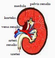
Human kidney has a length of about 10 cm and forms such as red beans, numbered pair, and is located on the left and right of the spine. Type metanefros the human kidney is not segmented and have many glomeruli. Kidneys are the skin of the kidney, kidney marrow, and kidney cavity.
  1. Skin the kidney (cortex). In the skin there are many kidney Malpighi bodies totaling ± 1 million. Agency consists of glomerular Malpighi.
  2. Kidney marrow (medulla). Kidney marrow bodies form a cone-shaped and contains a lot of channels that collect urine called convoluted tubules.
  3. Cavity kidney (renal pelvis). In this cavity empties into the collecting duct. Of the cavity, urinary tract out of the ureter to the bladder (bladder). From the bladder, urine exits the body through the urethra.
b. Formation of Urine in Kidney

The process of formation of urine in the kidneys through three stages as follows:

1) Filtration (filtering)Blood filtration occurs at the glomerulus, the blood capillary coiled inside the Bowman's capsule. In glomerular endothelial cells are thus facilitate the screening process. In addition, the binding also occurs in glomerular blood cells, platelets, and most of the plasma proteins that did not participate were excluded. The results of the infiltration process in the form of primary urine (glomerular filtrate) is compositionally similar to the blood, but does not contain protein. In the primary urine can be found amino acids, glucose, sodium, potassium, ions, and other salts.

2) Reabsorption (reabsorption)
The process of reabsorption occurs in the vessels (tubules) proximal. This process occurs after the primary results of the infiltration of urine flow in the vessels (tubules) proximal. The ingredients are absorbed in the process of reabsorption are materials that are still useful, such as glucose, amino acids, and a large number of inorganic ions. In addition, the water contained in the primary urine is also experiencing reabsorption by osmosis, while the other ingredients reabsorption take place in active transport.

The process of water absorption also occurs in the distal tubule. Then, the materials that have been reabsorbed by the proximal tubule is returned to the blood through the capillaries that surround the tubules. This reabsorption process also occurs in the loop of Henle, especially sodium ions. The result of the process of reabsorption is secondary urine composition constituent substances which are very different from the primary urine. In the secondary urine was not found substances that are still needed by the body and urine levels higher than in the primary urine.

3) Augmentation (Addition)Urine subsequent secondary distal convoluted tubule entry into and collecting ducts. This happens in the process of adding channels substances that do not benefit the rest of the body. Then, the actual urine get into the bladder (urinary bladder) through the ureter. Furthermore, the urine will be excreted from the body through the urethra. Urine contains urea, urine acid, ammonia, and disassembly of protein remnants. In addition, substances containing excessive in the blood, such as vitamin C, drugs, and hormones and salts.